Polska wersja |
Photos of
luminescent bacteria cells |
|||

|
|
|||||
![]()
![]() Some photos of luminescent bacteria cells:
Some photos of luminescent bacteria cells:
![]()
![]()
![]()
![]() There are some
photos of luminescent bacteria cells. Sorry, quality of these
photos is not good, but I do not have any special equipment. I
took the photos of stained cells. I used popular light microscope
and special adapter (photographic ring), which make me able to
join the microscope and my camera. The photos, which I got were
corrected by use of computer graphic programs to get better
sharpness and more true colours. I started four cultures of four
luminous bacteria species on the petri plates with "BOSS"
medium. I prepared two microscopic preparations from each culture
in two ways of stain (I used Gram procedure of staining and
simple negative staining procedure)
There are some
photos of luminescent bacteria cells. Sorry, quality of these
photos is not good, but I do not have any special equipment. I
took the photos of stained cells. I used popular light microscope
and special adapter (photographic ring), which make me able to
join the microscope and my camera. The photos, which I got were
corrected by use of computer graphic programs to get better
sharpness and more true colours. I started four cultures of four
luminous bacteria species on the petri plates with "BOSS"
medium. I prepared two microscopic preparations from each culture
in two ways of stain (I used Gram procedure of staining and
simple negative staining procedure)
1. |
2.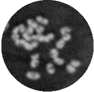 |
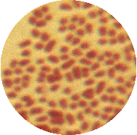
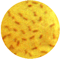
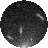
![]() Photobacterium phosphoreum (Vibrio
phosphoreum) - photo 1 shows Gram stained bacterial cells.
The shapes of them are cylindrical or almost spherical, Gram-negative.
Photo 2 shows negatively stained cells - more cylindrical cells
are visible. I think that the shapes of some of them are like the
bean seed
Photobacterium phosphoreum (Vibrio
phosphoreum) - photo 1 shows Gram stained bacterial cells.
The shapes of them are cylindrical or almost spherical, Gram-negative.
Photo 2 shows negatively stained cells - more cylindrical cells
are visible. I think that the shapes of some of them are like the
bean seed ![]() .
.
1. |
2.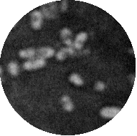 |
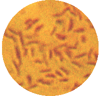
![]() Photobacterium
luciferum. Cylindrical, Gram-negative cells are visible.
Photo 1 shows Gram stained cells and photo 2 shows negative
stained cells of this bacterium.
Photobacterium
luciferum. Cylindrical, Gram-negative cells are visible.
Photo 1 shows Gram stained cells and photo 2 shows negative
stained cells of this bacterium.
1. |
2. |
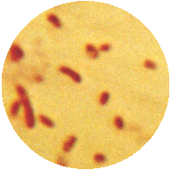
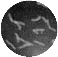
![]() Vibrio
fischeri. Photo 1 shows cylindrical cells of this Gram-negative
bacterium. Cylindrical shape is more visible when cells are
negative stained (photo 2).
Vibrio
fischeri. Photo 1 shows cylindrical cells of this Gram-negative
bacterium. Cylindrical shape is more visible when cells are
negative stained (photo 2).
1. |
2. |
![]() Vibrio
harveyi cells. Cells are cylindrical, Gram-negative (photo 1).
Cylindrical shape is more visible when cells are negative stained
(photo 2).
Vibrio
harveyi cells. Cells are cylindrical, Gram-negative (photo 1).
Cylindrical shape is more visible when cells are negative stained
(photo 2).
| HOME PAGE | NATURAL ENVIRONMENT | MORPHOLOGY AND PHYSIOLOGY |
| These pages were created by Piotr Madanecki |
| e-mail: pmad@eniac.farmacja.amg.gda.pl |